News & Blog
News & Blog
Press Release & Community Statement: PTC Therapeutics Receives Complete Response Letter for Vatiquinone NDA

August 2025 – Advocacy Newsletter
Take Action to Raise Awareness for FA—Submit a Proclamation Request!
Job Posting: Research Portfolio Manager
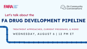
FA Community Conversations FA Drug Development Pipeline Webinar: Recording Available on YouTube
Design Therapeutics Highlights Progress Across Lead GeneTAC® Programs and Reports Second Quarter 2025 Financial Results
Meet the 2025 FARA Fellows
Press Release and Community Statement: Lexeo Therapeutics Announces FDA Breakthrough Therapy Designation for LX2006 in Friedreich Ataxia
June/July 2025 – Advocacy Newsletter
